, actin (red), vinculin (pink) and DNA (blue).")
Caco-2 cells labeled for tight junction molecule cingulin (green), actin (red), vinculin (pink) and DNA (blue).

Epithelial cells growing on a patterned adhesive surface with the shape of the Weizmann Institute tree.
.")
Desmosomes in mouse tongue epithelium (by transmission electron microscope).
 and phospho-tyrosine (red).")
Porcine aortic endothelial cell, double-labeled for actin (green) and phospho-tyrosine (red).

“Molecular composition map” of focal adhesions and stress fibers.
.")
Myeloma cancer cell responding to shear flow (by scanning electron microscope).
Nano-architecture of adhesion complexes
In the 1970's, electron microscopy of focal adhesions revealed the close (~15 nm) association of the plasma membrane with the underlying substrate, and multiple filaments, mostly actin microfilaments, running along the cytoplasmic faces of the ventral membrane (Figure 1).
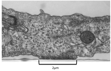
Figure 1
Electron microscopy of focal adhesions. Transmission electron microscope image of a chicken lens cultured cell. Black arrow indicates cell-surface adhesion. Image adapted from Medalia and Geiger, 2010.
Recently, “doughnut”-shaped particles with a typical diameter of ~25 nm, bridging between actin filaments and the membrane, were detected in that area. These novel integrin adhesion-associated complexes were found using correlated cryo-electron tomography and light microscopy (Figure 2),giving rise to the notion that focal adhesions are actually made of a small set of dynamic protein complexes. Utilizing biochemistry and mass spectrometry, several adhesion-related protein complexes were found (Figure 3).
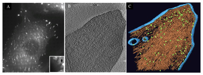
Figure 2
Cryo-electron tomography of integrin-mediated focal adhesions. (a) Correlated microscopy, combining fluorescence microscopy at room temperature (using a cell line expressing YFP–paxillin) and cryo-electron tomography. The area indicated by the brackets in A was visualized, following vitrification, using low-magnification cryo-electron microscopy (inset) and the individual focal adhesion, indicated by the yellow and red arrows, was subjected to cryo-electron tomography. (b) A 10 nm slice through a cryo-tomogram of the focal adhesion indicated in A shows aligned actin filaments and the plasma membrane. (c) Surface-rendering view of the focal adhesion site as seen from the direction of the substrate towards the cell. Actin is depicted in brown, membranes in blue and a large number of uniformly-oriented particles located at the interface between the cytoskeletal bundle and the membrane, which are probably adhesion-related particles, are depicted in green.

Figure 3
Isolation and identification of adhesion associated protein complexes. (a) Chicken gizzard smooth muscle extracts separated by gel filtration. Elution pattern suggests the existence of several protein complexes. (b) Mass spectrometry analysis of selected fractions indicates the existence of a vinculin-Arp2/3 hybrid complex. (c) According to results, Arp2/3 is a modular complex made of 2 modules. An actin nucleating module made of Arp3, Arp2, p34 and p21, and a localizing module composed of p41, p16 and p20, which can be exchanged with other proteins such as vinculin.
Further Reading
(2010).
Dissecting the molecular architecture of integrin adhesion sites by cryo-electron tomography.
Nature Cell Biology.
12
(9):909-915.

(2012).
Reconstructing adhesion structures in tissues by cryo-electron tomography of vitrified frozen sections.
Journal of Structural Biology.
178
(2):76-83.
(2014).
Regulation of focal adhesion formation by a vinculin-Arp2/3 hybrid complex.
Nature Communications.
5
:1-11.