
Following pruning, γ neurons undergo developmental axon regrowth. Recently, we have provided the first evidence that developmental axon regrowth is genetically regulated, distinct from initial axon outgrowth and shares similarities with axon regeneration following injury. By combining genetics and genomics, we aim to provide a deep mechanistic understanding of axon regrowth that should, in the long run, impinge on our understanding of the constraints of axon regeneration following injury.
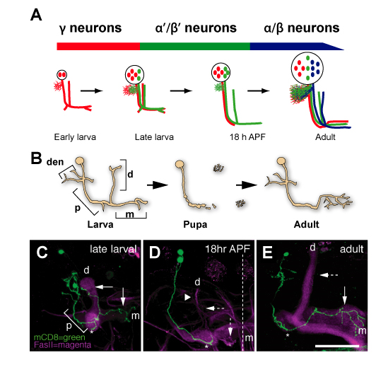 Figure 1: Neuronal remodeling of Drosophila Mushroom Body (MB) γ Neurons. (A) Schematic summary of MB development, indicating the three types of neurons generated from a common neuroblast precursor at three developmental stages. γ (red), α’/β’ (green) and α/β (blue) neurons have distinct axonal projection patterns in the adult brain. NHL, newly hatched larvae; ALH, after larval hatching; APF, after puparium formation.(B) Schematic representation and (C-D) γ neuron single/two-cell MARCM clones at different stages during development. (B,C) Larval neurons project axons to both the dorsal (d) and medial (m) lobes. (B,D) At 18 hr APF, axons prune larval-specific dorsal and medial branches (dashed arrows indicate the absence of the larval branches). (B,E) Adult neurons re-extended only medial (solid vertical arrow) but not dorsal (dashed horizontal arrow) branches. den, dendrites; p, peduncle; Green in (C-E) represents MB γ neuron MARCM clones labeled with mCD8-GFP; magenta represents anti-FasII staining, which labels strongly α/β neurons, weakly γ neurons, and does not label α’/β’ neurons. Modified from Watts et al (2003).